We understand how stressful it is choosing an imaging software. When researching imaging solutions, it’s important to compare several factors—product features being one of them. Dedicated to making your life easier, Apteryx Cloud Imaging aims to improve your practice by including essential features within your software.
- What imaging features do I need?
- How do the features affect productivity in my practice?
- How is Apteryx different from other imaging solutions in the market?
This blog will help answer your questions and provide clarity on the best imaging features and how they can benefit you, your practice, and your patients!
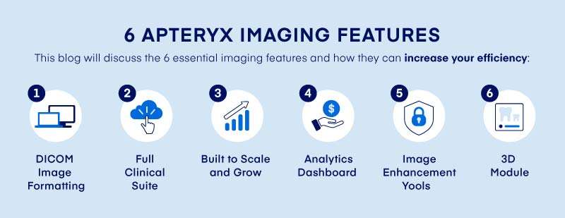
Apteryx offers the most desired features to help grow your practice while simplifying your workflow. Our all-in-one software is designed to include the top 6 essential features to increase your practice’s efficiency while increasing your competitive advantage over your competitors.
1. DICOM Image Formatting
DICOM (Digital Imaging and Communications in Medicine) has been the image format standard for the last 30 years. While it is a standard, many industries in healthcare—including dentistry—don’t always operate in this format, resulting in the loss of patient data.
DICOM – allows reliable transmission of information between the devices taking the images, devices storing the images, and devices displaying the images.
Apteryx Imaging not only imports DICOM formatting but can also store it as a DICOM file (.dcm) and easily export it. With DICOM, your data is readable from multiple types of software rather than just the one you are currently using for imaging. This is helpful in speeding up the process should you need to refer a patient. You also no longer need to convert proprietary format to a readable format when changing systems or acquiring a practice.
Advantages of DICOM image formatting include:
- Simple image transfer when changing systems and practices
- Freedom of imaging solution
- No need for complicated and expensive data conversions
- Easy image integration across digital imaging devices
DICOM ensures your patient’s image data is not only readable by most digital imaging devices (intraoral sensors, digital pan/ceph devices, etc.) but also that the creation, transfer, and storage of your image is entirely HIPAA compliant at every stage.
With DICOM, your data is attached to the image and easily viewable for any referrals to outside consults, hospitals, and for other surgical purposes.
2. Full Clinical Suite
Apteryx’s all-in-one imaging software includes a full clinical suite to meet your needs—from a variety of series to image tools, Apteryx has all the tools you need to operate successfully!
Series Include:
- Full mouth
- Bitewing
- Intra/extraoral camera
- Pano/ceph
These x-rays are provided in multiple angles to easily identify concerns while fulfilling your diagnosis needs.
Our Clinical Suite Includes:
- Basic editing tools
- Image enhancement tools
- Measurement tools
- Image comparison
- Labeling & annotation
- Templates
Along with these features, the clinical suite also includes information and communication tools to view information about the patient, user, device, and which computer with tools to import, export, and share the images.
We’re dedicated to helping you meet your needs by delivering a powerful all-in-one imaging software with a full clinical suite.
3. Analytics Dashboard
To maximize your practice’s efficiency, Apteryx Analytics Dashboard saves time by tracking and filtering your activities. This user-friendly dashboard can easily filter between locations, viewing all locations at once or a specific one.
Apteryx Analytics Dashboard Includes:
- Capture station activity
- Acquisition activity
- Device activity
- Operator activity
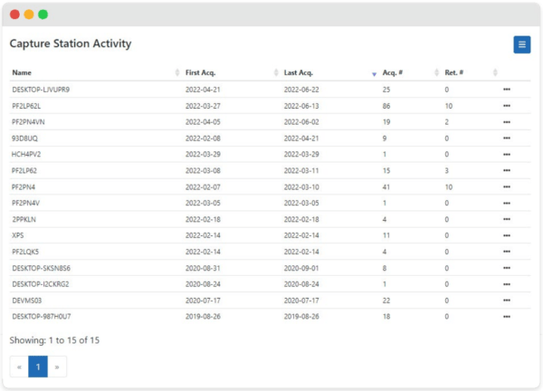
The Capture Station Activity displays valuable information on how many acquisitions were taken on a station and what the retake number is per capture station. This is especially helpful when wanting to see which computer may be having issues due to a high retake number.
Acquisition Activity displays information from acquisition and retakes in simple digestible visuals such as graphs and pie charts. You can also break it down by gender to track your activity. This creates an easy overall view of the analytical data within the dashboard over time
Device Activity shows the serial number of each device. This is especially useful when facing image quality issues. Looking at the serial number and number of retakes can help you identify which sensor is having difficulty, so it can be fixed. Another benefit is comparing acquisition numbers of each device to see which brand lasts longer.
Operator Activity shows who took the first and last acquisition. Using the name and number of retakes, you can easily view which employees are doing well or having troubles and need additional assistance. This helps identify the source of the problem for a quick resolution.
4. Image Enhancement Tools
In addition to basic editing tools (zoom, center, rotate, and flip), these 9 image enhancement tools are guaranteed to help you visualize and analyze your images with ease and clarity.
Whether you are looking to enhance a single image or a series, our tools will help clarify your image to easily diagnose your patient and identify concerns.
Apteryx Image Enhancement Tools:
- Exposure
- Brightness
- Contrast
- Gamma
- Denoise
- Spot enhancement
- Enhancement tool
- Filters
- Sharpness
These enhancement tools can also be saved and reset to save time by avoiding starting from scratch and making manual adjustments.
5. Secure Sharing Portal
Do you need to securely share images with 3rd parties for referrals or consulting? Apteryx has got you covered!
Our secure sharing portal offers your patients more comprehensive care by enabling the sharing images to other essential figures through the cloud. Examples of 3rd parties include providers, external laboratories, and insurance companies.
Benefits of Secure Sharing Portal Include:
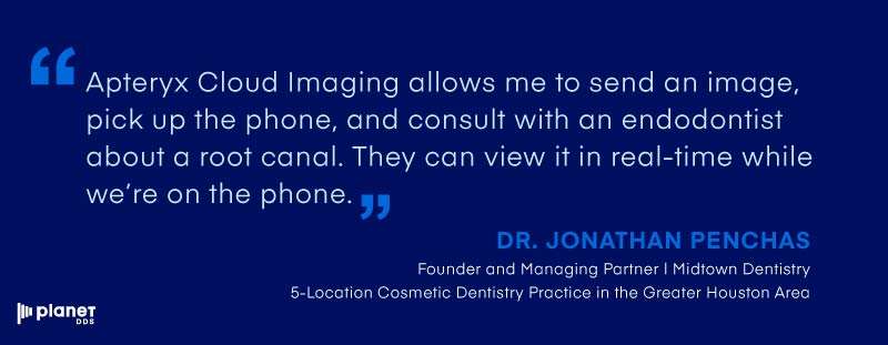
- Consult external parties
- Real-time decisions
- HIPAA-compliant portal
Now, you can share images with external parties by sharing clinical data through a secure HIPAA-compliant image portal to provide seamless opportunities for collaboration and referrals.
6. 3D Module
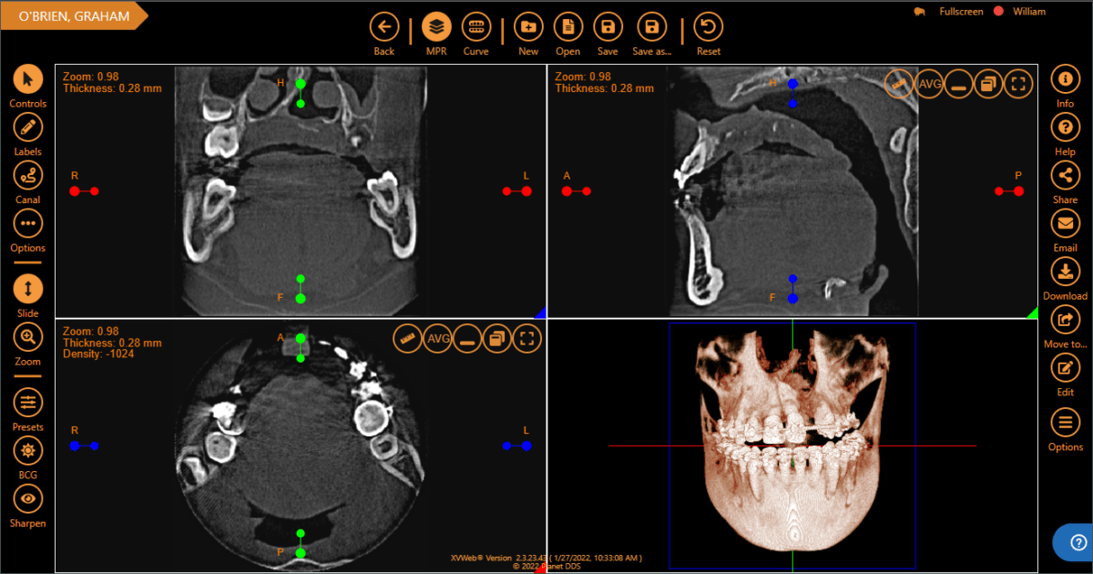
If your practice utilizes 3D imaging like CBCT (cone-beam computed tomography) scans, finding an imaging software with 3D imaging capabilities perfect for you and your practice is crucial.
Apteryx’s 3D module allows you to securely access and share CBCT and STL (stereolithography) datasets online. STL files help share 3D images without formatting your data to 2D slices.
Stereolithography—Describes surface geometry of a three-dimensional object
With dental practices offering dental implant treatments and other oral surgeries, 3D imaging is very common and necessary to fully understand your patient and provide the best treatment for them.
How 3D Imaging Helps You:
- Easier problem detection
- Oral surgery
- Dental implant treatments
- Scanning the mouth
What makes the 3D module different from the 2D clinical suite? Along with all the basic tools and image enhancement tools of the clinical suite, 3D has a more advanced and comprehensive toolset designed to help you reach your practice’s potential and increase efficiency.
3D Module Features Include:
- Curved slice/pano reconstruction
- Implant Planning
- Volume Rendering
- Nerve Canal Tracing
- Extensive Implant Library
- Effects Presets
- Treatment Planning
- Effects Presets
Apteryx securely accesses and shares 3D images without needing to download large files while providing a wide array of visualization and diagnostic capabilities for CBCT datasets.
These 6 essential features are designed to optimize your practice’s workflow by increasing productivity and efficiency. Join the future today and boost your practice with Apteryx Cloud Imaging.
Missed Part 1? Check It Out Now: Top 5 Benefits of Apteryx – Part 1: Why Apteryx Series

